Cart 0

Research Areas

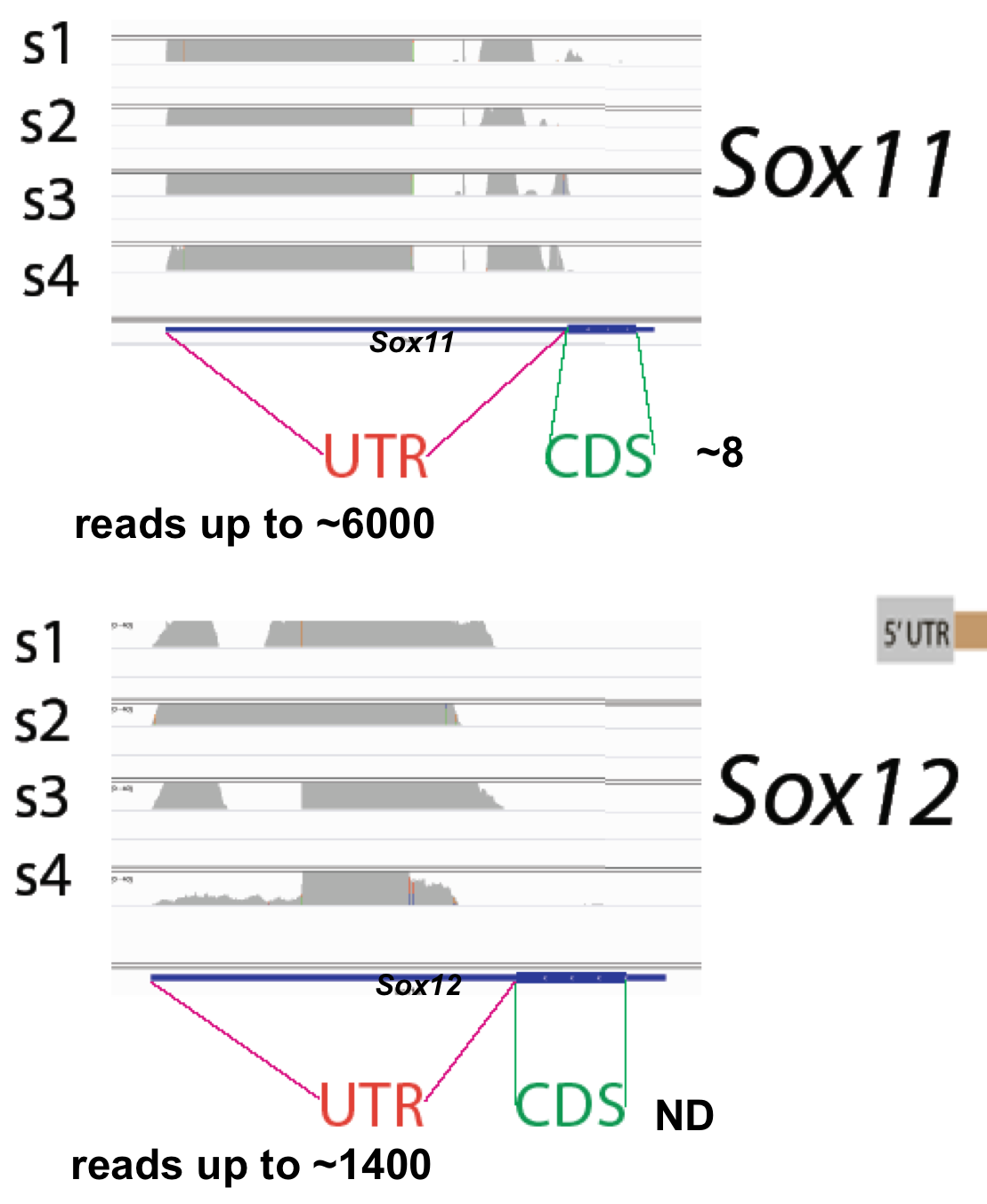
In each paradigm we focus on understanding the role of isolated 3'UTRs, 3'UTR sequences expressed in the absence of their cognate coding regions (3'UTR shown in red, CDS in green and protein in blue, in pictures below)

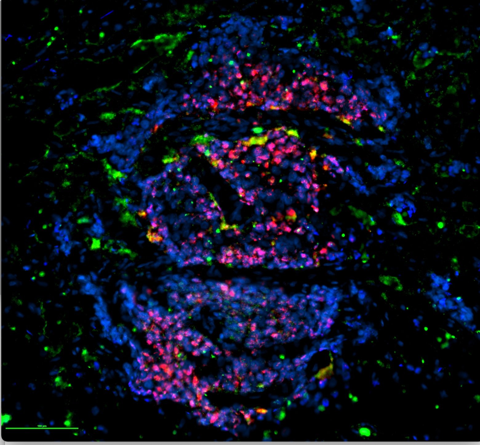
Pluripotent gene 3’UTR (red), cognate CDS (green), in human embryonic stem cell 2D organoid

Embryonic Stem cells

We are focused on the role of pluripotent gene i3’UTR and CDS in stem cell organization, proliferation and quiescence using bioinformatics, Crispr and other approaches.

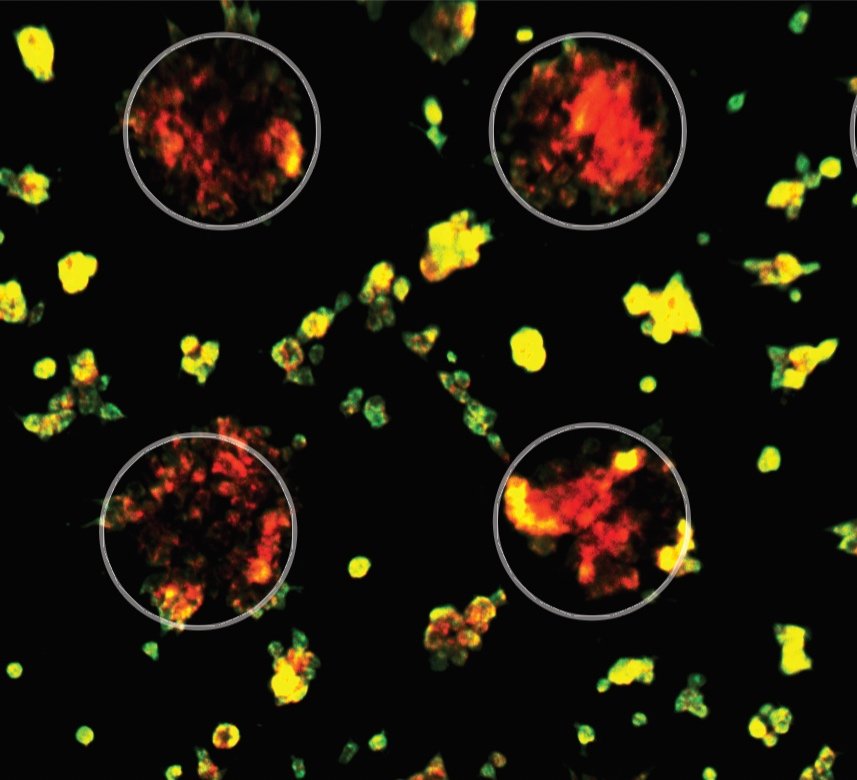
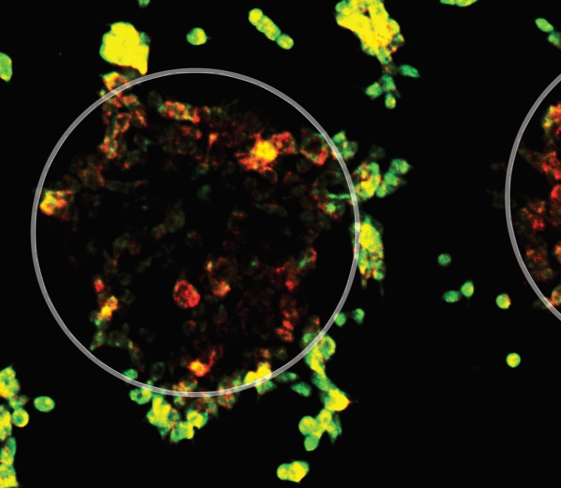
Cancer gene 3’UTR (red), cognate CDS (green), in human cancer cell line. Aggregated cells (in 2D organoid circles) express high 3’UTR, migrating cells switch to high CDS for the same gene.

Cancer

We are investigating the role of isolated 3’UTR expression in cancer progression, using bioinformatic and Crispr approaches in cancer cell lines, human cancer tissue and in vivo.


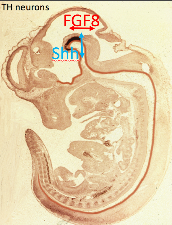
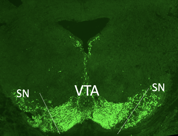
Developing mouse dopamine neurons (blue), transiently express the Nurr1 CDS (green), then switch back to Nurr1 3’UTR expression (red). Dorsal is up.

Neuronal development

Our lab has a long standing interest in developing dopaminergic neurons. Currently we are examining the role of i3'UTRs in the cell fate specification of developing dopamine neurons. 

Embryonic stem cells 

We use 2 and 3D organoids, as well as harvested blastocysts to study the role of the differential 3’UTR and CDS expression of pluiripotent genes in stem cell proliferation and quiescence, using a combination of over-expression and Crispr knockdown, together with bioinformatic analyses.  

Screen Shot 2022-02-14 at 8.44.13 AM.png

Cancer

(Left, center)cells in culture showing 3’UTR (red) CDS (green) for gene 1. (Right) breast cancer tissue; 3’UTR (red), CDS (green) for gene 2.


Neuronal Development

A long-term focus of our laboratory has been the investigation of developing dopaminergic neurons (DA neurons), the key cells that die in Parkinson’s disease (left two panels). Originally we and colleagues were the first to show that dopaminergic neurons are induced by the co-presentation of two molecules Sonic hedgehog and FGF8. This work has led to a widely used method involving a combination of these and other molecules to derive dopaminergic neurons from embryonic stem cells.

We are currently examining how the differential expression of 3'UTR/CDS sequences, first identified by us in developing DA neurons (Kocabas et al 2015; Neuron) might play a role in dopaminergic and other neural differentiation or in disease related events.  We find that for key genes related to DA neuron development there are age related changes in 3'UTR/CDS ratios within these neurons.  A combination of in vitro, in vivo and bioinformatic approaches is being used to investigate these processes.  

Sox C genes

Another project in the lab is to investigate the role of Sox C genes (Sox4, 11, and 12) in neuronal development, these genes are highly expressed in the brain and are important developmental regulatory genes.  Traditional in situ hybridization using a single probe to the Sox11 mRNA shows widespread, almost ubiquitous expression in the developing brain (bottom). However two color in situ hybridization of Sox11 reveals exquisite patterns, gradients and cell type specific expression within the developing nervous system (top), suggestive of important functions for this differential expression. Viral overexpression of 3'UTR sequences and examination of Sox C knockout mice will allow us to test these predictions. 

Sox 11 3'UTR (red) CDS (green) in E15 brain and region of developing Dopaminergic neurons (white arrow)

Sox 11 3'UTR (red) CDS (green) in E15 brain and region of developing Dopaminergic neurons (white arrow)

Sox family members in developing eye show different patterns;. 3'UTR (red), cognate CDS (green). 

Sox family members in developing eye show different patterns;. 3'UTR (red), cognate CDS (green). 

Left Sox11 in situ using two color in situ hybridization with a red probe to the 3'UTR and green probe to the CDS; note dramatic layered patterns of Sox 11 3'UTR vs CDS in developing cortex.  Right single color in situ of Sox11 CDS from Al…

Left Sox11 in situ using two color in situ hybridization with a red probe to the 3'UTR and green probe to the CDS; note dramatic layered patterns of Sox 11 3'UTR vs CDS in developing cortex.  Right single color in situ of Sox11 CDS from Allen Brain Atlas. (http://developingmouse .brainmap.org/experiment/ show/ 100071526.) 

Screen Shot 2018-02-23 at 11.04.14 AM.png